
Choosing a Breast Size for Reconstruction Starts With Your Body and Your Goals
When choosing breast size for reconstruction, start with the look you want. Then ask your surgeon to measure your chest and assess the skin and tissue available after mastectomy. The best size is one that fits your frame, supports your goals, and does not place too much strain on healing tissue. Implant volume alone cannot tell you how the result will look.
Your original breast size, whether you are rebuilding one or both breasts, and any plans for radiation also matter. If your reconstruction uses a tissue expander, its gradual fills may help you and your surgeon judge the size before choosing a permanent implant. Understanding the full scope of breast reconstruction can help you feel more prepared for these decisions.
I’m Dr. John A. Cece, a plastic and reconstructive surgeon with fellowship training in aesthetic surgery and microsurgical breast reconstruction. My experience with implant-based and flap procedures shapes how I approach choosing breast size for reconstruction: as a decision we make together, with your goals and safety in mind.

Clinical and Anatomical Factors in Choosing Breast Size for Reconstruction
Selecting the right volume involves far more than picking a cup size off a shelf. In aesthetic breast augmentation, the native breast tissue and glandular structure remain intact to provide natural padding. In post-mastectomy reconstructive breast surgery, the oncologic surgeon removes the glandular breast tissue, leaving behind a delicate skin envelope that requires careful management.
Whether your reconstruction is placed prepectorally (above the chest muscle) or subpectorally (beneath the pectoralis major muscle), the quality and vascular supply of your mastectomy skin flaps dictate how much volume the tissue can safely support. During your initial consultation, we perform a pinch test to measure the thickness of the remaining subcutaneous tissue. A thicker soft-tissue envelope allows for greater flexibility with implant dimensions, whereas thinner skin demands careful implant selection to prevent visibility, rippling, or circulatory stress.
Tissue-Based Measurements When Choosing Breast Size for Reconstruction
To achieve a balanced contour that harmonizes with your chest wall, we rely on precise anatomical landmarks:
- Breast Base Width (BBW): The horizontal diameter of the breast footprint on your chest wall. This is the single most critical measurement because an implant that exceeds your natural base width will extend into the axilla (armpit) or cross the sternal midline.
- Sternal Notch-to-Nipple Distance: Establishes the vertical proportion of the breast mound on your torso.
- Inframammary Fold (IMF) Position: The natural lower crease where the breast meets the chest wall, determining the lower pole projection.
- Intermammary Distance: The separation between your breasts, ensuring natural cleavage without medial touching (symmastia).
By measuring these dimensions, we can match your body’s natural frame to the exact width and projection profiles in implant-based breast reconstruction, ensuring the new breast sits symmetrically on your chest wall.
How Mastectomy Specimen Weight and Density Influence Volume
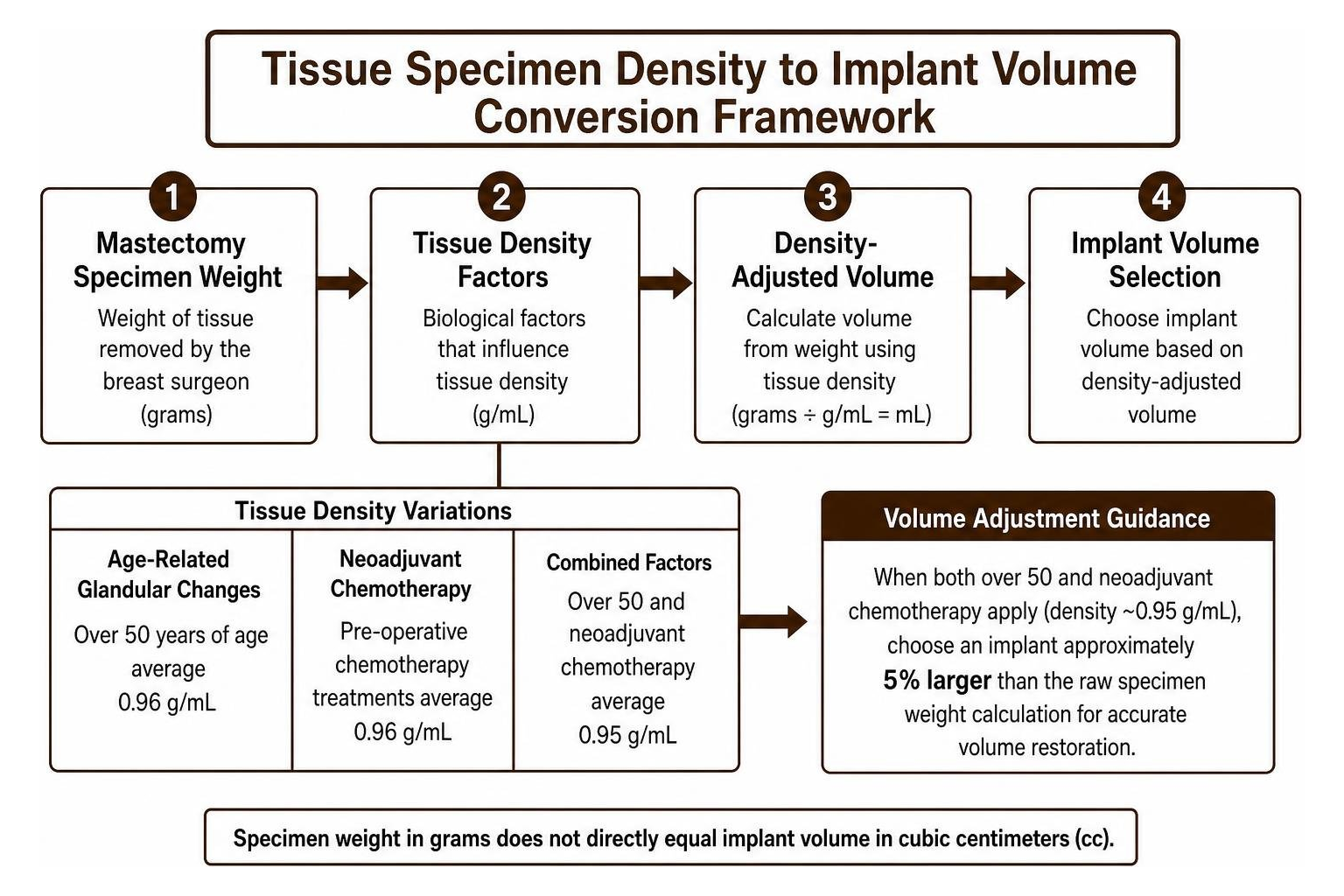
Patients often expect a 1:1 match between the weight of tissue removed by the breast surgeon and the volume of the implant placed. However, clinical studies show that specimen weight in grams does not directly equal implant volume in cubic centimeters (cc) Factors affecting mastectomy specimen density in direct-to-implant breast reconstruction .
Breast tissue density varies significantly based on biological factors:
- Age-Related Glandular Changes: Patients over 50 years of age typically exhibit lower tissue density (averaging 0.96 g/mL compared to 1.01 g/mL in younger patients), meaning their tissue is lighter per unit of volume due to higher fatty replacement.
- Neoadjuvant Chemotherapy: Pre-operative chemotherapy treatments alter glandular density, reducing it to approximately 0.96 g/mL.
- Volume Adjustments: When a patient is over 50 and has completed neoadjuvant chemotherapy, combined density drops to roughly 0.95 g/mL. In these cases, choosing an implant approximately 5% larger than the raw specimen weight calculation provides accurate volume restoration.
Selecting Implant Profiles, Dimensions, and Fill Materials

Once we identify your anatomical base width, we determine the forward projection and fill characteristics of the prosthetic device. Two implants with the exact same volume (such as 350cc) can create entirely different silhouettes depending on their base diameter and forward projection height.
Low, Moderate, and High Profile Projection Comparison
Implant profile refers to how far forward the implant projects from the chest wall relative to its base width:
- Low Profile: Features a broader base width with minimal forward projection. This is ideal for patients with wide chest dimensions who desire subtle fullness without dramatic forward protrusion.
- Moderate and Moderate Plus Profile: Offers a balanced ratio of base diameter to projection. This profile provides natural slope contouring and represents the most versatile choice for standard body frames.
- High and Ultra-High Profile: Features a narrow base diameter combined with maximum forward projection. This configuration is essential for patients with narrow chest walls who require adequate projection without the implant extending laterally past the rib cage.
| Profile Type | Base Width Characteristic | Forward Projection | Ideal Patient Anatomy |
|---|---|---|---|
| Low Profile | Wide | Subtle / Flatter | Broad chest wall, shallow anterior projection goals |
| Moderate Profile | Balanced | Natural Curve | Average torso proportions, natural anatomical slope |
| High Profile | Narrow | Pronounced | Petite or narrow chest wall needing focal projection |
Silicone Gel vs. Saline Fill Dynamics
The choice between saline and silicone fill materials affects both aesthetics and surgical planning:
- Highly Cohesive Silicone Gel (“Gummy Bear”): Form-stable silicone implants utilize cross-linked cohesive gel that maintains upper-pole fullness and resists collapsing or wrinkling. For prepectoral reconstruction, silicone and cohesive gel implants are frequently preferred because they significantly minimize visible edge rippling under thin mastectomy skin. Silicone devices come pre-filled, requiring a slightly longer incision for placement.
- Saline Devices: Filled with sterile saline solution during surgery through an empty silicone elastomer shell, allowing insertion through smaller surgical incisions. However, in reconstructive settings with thin skin flaps, saline can carry a higher incidence of palpable rippling or upper pole scalloping unless supported by autologous fat grafting.
Tissue Expander Sizing and Staged Reconstruction Protocols

When direct-to-implant placement is not clinically recommended—such as when the mastectomy skin flaps require time to stabilize—two-stage expander-to-implant reconstruction offers a controlled approach. In this technique, a temporary expander is placed under the skin or muscle and supported with acellular dermal matrix (ADM).
Using the Expander as an In Vivo Sizer
One advantage of staged reconstruction is that the tissue expander serves as a real-time sizing tool. After an initial healing period of two to four weeks, we begin gradual serial expansions in our office. This allows you to experience the physical weight, visual proportion, and wardrobe fit of different volumes. If you initially planned for a 350cc reconstruction but find that 450cc provides better symmetry with your natural side, the expansion volume can be tailored accordingly before selecting your permanent implant or exploring supplementary autologous breast reconstruction techniques.
Expansion Velocity, Volume Limits, and Revision Thresholds
Clinical research shows that expansion protocols directly impact complication rates and final outcomes (Kim et al., 2022). In studies evaluating expansion strategies, bi-weekly filling increments of 50cc to 100cc that maintain steady expansion velocity help minimize dead space around the device and reduce seroma formation (Kim et al., 2022).
To ensure long-term tissue safety, expanders are typically filled to between 60% and 80% of capacity before exchange, with final expansion volumes capped at or below 120% of rated capacity to prevent excessive skin blanching and vascular strain The investigation of the relation between expansion strategy and outcomes of two-stage expander-implant breast reconstruction.
Safety Considerations and Complication Risks by Breast Size
While achieving your aesthetic goals is essential, surgical safety remains our top priority. Selecting an implant that exceeds the vascular carrying capacity of your skin flap significantly elevates postoperative risks.
Key Risks and Trade-Offs in Choosing a Breast Size for Reconstruction
Published clinical data indicates that reconstructed breast volume directly correlates with postoperative complications:
- Infection and Prosthetic Failure: Multivariate analyses show that large breast size (specimen weight exceeding 500 grams) is a significant risk factor for complications, increasing the odds ratio to 3.20 (Suga et al., 2017). In large-volume reconstructions (>500g), infection rates reach approximately 7.01% and expander failure reaches 5.26%, compared to 1.52% in medium-sized breasts (300g to 500g) Risk Factors for Complications in Expander-Based Breast Reconstruction: Multivariate Analysis in Asian Patients.
- Mastectomy Type and Flap Necrosis: Nipple-sparing mastectomy carries a higher incidence of skin flap ischemia compared to skin-sparing or total mastectomy due to the extended vascular path required to sustain the nipple-areolar complex.
- Radiation Therapy: Irradiated tissue loses elasticity and vascularity, necessitating conservative sizing parameters or secondary revision support through breast revision surgery.
Quantitative Revision Cut-Offs and Safety Guidelines
Receiver Operating Characteristic (ROC) statistical modeling highlights specific safety thresholds during reconstruction planning:
- Weight Cut-Off: A mastectomy specimen weight exceeding 696 grams predicts an increased likelihood of revision surgery, with a specificity of 81.8% (Kim et al., 2022).
- Volume Cut-Off: A reconstructed volume exceeding 505cc yields a sensitivity of 64.3% and specificity of 66.2% for requiring secondary corrective procedures.
When a patient presents with larger native breasts (>700g), achieving safe symmetry often involves choosing a moderate-sized implant (300cc to 450cc) for the reconstructed breast while performing a contralateral balancing procedure, such as a breast reduction or mastopexy (breast lift), on the remaining breast.
Frequently Asked Questions About Reconstruction Sizing
How many cc’s are typical for a reconstructed breast implant?
Most reconstructive breast implants range between 200cc and 600cc. Volumes between 300cc and 450cc are the most common, providing balanced proportion for average anatomical chest widths while keeping tissue tension within safe physiological parameters.
Can I choose to be larger or smaller than my natural pre-mastectomy size?
Yes. Breast reconstruction offers the opportunity to adjust your baseline size. If you previously experienced neck or back strain from overly large breasts, you can choose to downsize during reconstruction. If you undergo a unilateral (single-side) mastectomy and wish to change your overall size, we can perform a reduction, lift, or augmentation on your contralateral (opposite) breast to establish symmetry.
Why does implant volume in cc differ from specimen weight in grams?
Implant volume measures liquid displacement (cubic centimeters), whereas specimen weight measures mass (grams). Because breast tissue consists of a variable mix of fibrous glandular tissue (higher density) and adipose fat (lower density), 400 grams of breast tissue does not equal 400cc of implant fill. Factors such as age, body mass index, and prior chemotherapy treatments shift this ratio, requiring precise surgical adjustments.
Partnering on Your Reconstruction Journey
Choosing the right breast size for reconstruction is a collaborative process that balances your personal aesthetic vision with anatomical safety. In our state-of-the-art facility in Montclair, New Jersey, our surgical team—led by board-certified plastic surgeons Dr. Allen D. Rosen M.D. and Dr. Valerie J. Ablaza M.D., alongside Dr. John A. Cece, fellowship trained and board eligible—delivers personalized surgical care designed around your comfort and health.
We invite you to schedule a comprehensive consultation with our team to explore our advanced breast reconstruction services and develop a reconstruction plan tailored to your body.


