
Nipple Inversion: What It Means and What to Do
Nipple inversion means the nipple turns inward instead of pointing outward. If it has always looked that way, it is usually harmless and may not need treatment. A nipple that newly turns inward should be checked by a healthcare professional, especially if the change affects one side or comes with a lump, discharge, or skin changes.
The cause guides the next step. Mild inversion may respond to gentle suction or, when breastfeeding, help with latching. Surgery is an option for people who want a lasting change, but the technique matters if future breastfeeding is important.
I am Valerie J. Ablaza M.D., a board-certified plastic surgeon at The Plastic Surgery Group of New Jersey in Montclair. My training in breast and aesthetic surgery informs my balanced approach to nipple inversion: first understand the cause, then weigh the options.

Understanding Nipple Inversion: Prevalence and Primary Causes
Nipple inversion is a widespread anatomical variation, affecting an estimated 2% to 10% of women, with broader population surveys showing that flat or inverted nipples occur in up to 10% to 20% of men and women alike. Anatomically, a typical nipple projects approximately 0.9 cm outward from the surrounding areolar tissue. In an inverted state, the nipple rests flush with or retracts below the plane of the areola.
The core mechanism behind this retraction involves the lactiferous (milk) ducts and the surrounding fibromuscular stroma. Beneath the areola, a web of smooth muscle fibers and collagenous connective bands anchors the skin to the deeper parenchymal tissue. When these connective bands are unusually taut, or when the milk-transporting ducts fail to lengthen during development, a mechanical tether is created that pulls the nipple inward.
Patients seeking care often want to understand the causes, symptoms, and treatment options for retracted and inverted nipples. As outlined by the National Cancer Institute definition of a retracted nipple, the condition can exist from birth or develop later in life secondary to trauma, inflammation, aging, or underlying tissue alterations. The psychological impact can be significant, often causing body image self-consciousness or anxiety regarding intimacy and nursing. Understanding the root etiology is the essential first step in determining the right clinical approach.
Congenital Factors and Anatomy
Congenital nipple inversion originates during fetal development. Around the sixth month of gestation, the mammary pit typically undergoes an eversion process where the underlying mesenchyme proliferates, pushing the nipple-areolar complex outward. When this mesenchymal proliferation is incomplete or tethered by dense connective tissue, the nipple remains invaginated.
Those exploring why congenital nipple inversion occurs from birth will find that approximately 90% of congenital cases are bilateral, affecting both breasts symmetrically. In a large systematic review of 3,369 cases, 93.9% of presenting inversions were congenital. These cases are characterized by shortened lactiferous ducts and rigid stromal bands that anchor the apex of the nipple to the underlying breast tissue, preventing natural projection without manual or mechanical traction.
Acquired Causes and Clinical Differences
Acquired nipple inversion accounts for roughly 6.1% of cases and develops after normal outward projection has already been established. Unlike congenital presentations, acquired inversion warrants immediate, detailed investigation because it is driven by secondary anatomical changes.
Common benign causes include duct ectasia—a condition where milk ducts become widened, clogged, and inflamed, leading to periductal fibrosis and shortening. Patients may also experience nipple shape changes during pregnancy as hormonal surges alter ductal architecture, or notice nipple retraction after pregnancy due to glandular remodeling and weaning. Other acquired causes encompass periareolar infections (mastitis or subareolar abscesses), post-surgical scar contraction following reductions or biopsies, significant weight loss, and natural age-related tissue atrophy. A detailed medical assessment, such as the UpToDate clinical overview of nipple inversion, emphasizes differentiating these benign, progressive conditions from acute pathological processes.
Clinical Grading: How Severity Is Evaluated

To standardize assessment and determine the appropriate corrective strategy, plastic surgeons rely on the clinically validated Han-Hong classification system (Han & Hong, 1999). This grading scale evaluates the degree of inversion, the presence of fibrotic tethering, the ease of manual extraction (the “pinch test”), and the structural integrity of the milk ducts.
| Feature | Grade 1 (Mild) | Grade 2 (Moderate) | Grade 3 (Severe) |
|---|---|---|---|
| Projection Mechanism | Easily pulled out manually or with cold/touch | Pulled out with moderate effort; retracts immediately | Difficult or impossible to pull out manually |
| Tissue Resistance | Minimal or soft fibrotic bands | Moderate fibrous tethering beneath the areola | Dense, rigid stromal fibrosis and severely shortened ducts |
| Maintenance of Eversion | Maintains projection for a period without traction | Inverts immediately once manual pressure is released | Remains completely retracted beneath the areolar plane |
| Lactiferous Duct Status | Ducts are normal length; minimal compromise | Ducts are mildly shortened but intact | Ducts are significantly shortened, tethered, or underdeveloped |
| Breastfeeding Potential | High; lactation is rarely impeded | Variable; may require latch assistance or pumps | Low without surgical duct preservation; often obstructed |
The Three Grades of Nipple Inversion
- Grade 1 (Mild): The nipple is easily pulled out with gentle manual pressure or responds directly to cold stimulation. Once everted, it maintains its projection for some time without immediately snapping back. Histologically, there is minimal stromal fibrosis, and the lactiferous ducts are fully developed without significant tethering.
- Grade 2 (Moderate): The nipple can be pulled out, but doing so requires notable effort. As soon as the physical pressure is released, it promptly retracts inward. Examination reveals moderate fibrotic bands anchored beneath the areola. Grade 2 is the most frequently encountered presentation in clinical practice, representing roughly 60% of cases evaluated in surgical settings as noted in foundational grading research published in Plastic and Reconstructive Surgery (Han & Hong, 1999).
- Grade 3 (Severe): The nipple is severely retracted and cannot be drawn outward during examination. The underlying tissue exhibits significant stromal deficiency, dense collagenous scarring, and markedly shortened ducts that rigidly bind the nipple to the underlying chest wall structures.
When Inversion Signals Breast Cancer or Infection
While lifelong inversion is generally benign, acute, newly acquired nipple inversion—particularly if unilateral—is a recognized clinical red flag. An underlying malignancy, such as invasive ductal carcinoma or ductal carcinoma in situ (DCIS), can incite a desmoplastic reaction (fibrous tissue growth) that contracts and pulls the retroareolar ducts inward. Paget disease of the breast, which accounts for 1% to 4% of breast cancer cases, frequently presents with nipple crusting, ulceration, scaling, and retraction.
In clinical studies of patients presenting with new-onset nipple inversion, between 5% and 6.5% were found to have an ipsilateral malignancy upon diagnostic imaging. Immediate evaluation is critical if inversion is accompanied by palpable lumps, skin tethering or dimpling (peau d’orange), or when nipple inversion occurs alongside spontaneous fluid discharge. Clinicians follow structured diagnostic protocols, such as the NHS clinical guidance for nipple problems, to rule out malignant disease before entertaining elective corrective procedures.
Diagnostic Evaluation and Imaging Protocols
A thorough diagnostic work-up begins with a detailed medical history and a comprehensive physical examination. We carefully determine the duration of the inversion, whether the onset was acute or progressive, and whether it is unilateral or bilateral.
During the physical examination, the surgeon or breast specialist assesses the skin of the nipple-areolar complex for eczema-like changes, erythema, or scaling. Palpation of the subareolar tissue and regional axillary lymph nodes is performed to check for discrete masses or adenopathy. The pinch test is applied to determine the structural grade and assess ductal tethering.

Diagnostic Mammography, Ultrasound, and When MRI Is Needed
For any patient presenting with new, acquired, or asymmetrical nipple inversion, formal breast imaging is the standard of care:
- Diagnostic Mammography: Bilateral diagnostic mammograms (including specialized craniocaudal, mediolateral oblique, and spot-compression retroareolar views) evaluate for suspicious microcalcifications, architectural distortion, or focal densities behind the nipple.
- Targeted Ultrasound: Targeted high-frequency sonography is routinely paired with mammography. Ultrasound is exceptionally effective at visualizing the subareolar ducts, differentiating solid masses from benign fluid collections (such as duct ectasia or cysts), and assessing ductal walls for intraductal papillomas. Together, mammography and ultrasound offer very high sensitivity and negative predictive value.
- Breast MRI and Biopsy: If physical examination and initial imaging reveal ambiguous retroareolar architectural distortion or if high clinical suspicion persists despite negative standard imaging, a contrast-enhanced breast MRI is indicated. If an identifiable mass or suspicious intraductal lesion is discovered, an image-guided core needle biopsy or skin punch biopsy of the areola is performed to definitively rule out malignancy before any aesthetic or reconstructive intervention.
Non-Surgical Solutions and Breastfeeding Strategies
For individuals with mild to moderate congenital inversion (Grade 1 and select Grade 2 cases), non-surgical management offers practical improvements for comfort and functional nursing. Addressing these concerns is equally important for women managing postpartum nipple changes when not breastfeeding, as hormonal transitions can alter tissue tone and baseline projection.
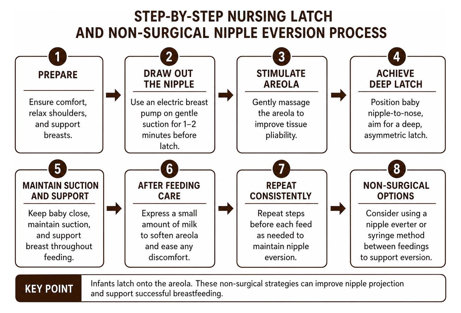
Managing Nipple Inversion for Lactation
Breastfeeding with inverted nipples is often entirely feasible, as infants latch onto the areolar tissue rather than solely the nipple tip. Successful nursing relies on increasing areolar pliability and drawing out the nipple tissue immediately prior to feeding:
- Pumping Before Latch: Utilizing a hospital-grade electric breast pump on a gentle suction setting for 1 to 2 minutes before nursing creates negative pressure that draws the inverted nipple outward, providing an accessible target for the newborn.
- Nipple Shields: Thin, food-grade silicone nipple shields can provide a firm, projected shape that stimulates the infant’s palate and encourages active sucking while milk transfer occurs.
- Lactation Guidance: Working closely with a certified lactation consultant helps optimize nursing positioning (such as the football or cross-cradle hold) to ensure the infant takes a deep, asymmetrical latch that encompasses ample breast tissue beneath the areola.
Manual Exercises and Suction Devices
Non-surgical tissue manipulation techniques aim to gently elongate the fibrous bands over time:
- Hoffman Technique: The application of the Hoffman technique involves placing the thumbs horizontally at the base of the areola and gently pulling outward in opposite directions, followed by repeating the motion vertically. Practiced consistently, this manual exercise helps stretch underlying adhesions.
- Suction Cups and Extractors: Non-invasive vacuum devices apply gentle suction over the nipple for designated daily intervals. While syringe extractors and suction cups can temporarily draw the nipple out—making them valuable tools right before infant latching—their ability to deliver permanent, long-term structural correction for Grade 2 and 3 inversions remains limited due to the natural elastic recoil of dense connective tissue.
Surgical Correction Techniques, Recovery, and Outcomes
When non-surgical measures prove insufficient, or when patients seek a definitive, long-term solution for aesthetic or functional concerns, surgical correction provides reliable, lasting projection. Inverted nipple correction is an outpatient procedure typically performed under local anesthesia, taking under an hour per side.
Patients interested in learning what to expect can review before-and-after results of nipple correction procedures to visualize realistic outcomes. The surgical literature confirms high success; an extensive systematic review of surgical correction of inverted nipples analyzing 3,369 cases demonstrated an overall satisfactory correction rate of 88.6%.
Duct-Preserving vs. Duct-Releasing Methods
The surgical strategy is selected based on the severity grade and the patient’s desire for future lactation:
- Duct-Preserving Techniques: Preferred for Grade 1 and Grade 2 cases where the patient wishes to preserve breastfeeding capability. The surgeon uses precise microdissection under magnification to release the tight fibrous bands surrounding the lactiferous ducts without transecting the ducts themselves. To maintain projection, internal tissue flaps—such as deepithelialized triangular dermal flaps or subcutaneous turn-over flaps (Jeong & Lee, 2015)—are transposed beneath the base of the nipple. This creates a supportive “tent-pole” or scaffold that bolsters the nipple and prevents it from sinking back into the breast stroma.
- Duct-Releasing (Transection) Techniques: For severe Grade 3 inversions characterized by rigid, severely shortened ducts, duct preservation may prevent adequate projection or lead to rapid recurrence. In these instances, the constricting ducts and fibrous cords are completely divided. A buried purse-string suture is then secured at the base of the areola to narrow the dermal ring and lock the nipple in an everted position. While this approach provides durable projection for severe cases, it eliminates future breastfeeding capacity from the treated breast.
Expected Results, Sensation, and Recurrence Rates
Following surgery, a protective dressing (often a custom “donut” or syringe-casing splint) is worn over the nipple-areolar complex for several weeks to prevent external compression from bras and clothing while internal healing occurs. Postoperative discomfort is generally mild and easily managed with over-the-counter or mild oral pain medications.
- Recurrence Rates: Modern surgical techniques demonstrate low recurrence rates, averaging approximately 3.89% across clinical studies. The use of vascularized subcutaneous flaps provides a substantial mechanical barrier against inward retraction compared to older, suture-only methods.
- Sensory Outcomes: Transient numbness or hypersensitivity is common during the initial weeks of healing. Because microdissection preserves the surrounding neurovascular bundles supplying the nipple-areolar complex, normal tactile sensation is preserved in the vast majority of patients once swelling subsides and nerve endings recover.
- Comprehensive Refinement: Nipple inversion correction can be performed independently or alongside broader aesthetic breast procedures, including female nipple reduction and reshaping or specialized male nipple correction approaches for symmetry, volume, and contour balance.
Frequently Asked Questions
Can inverted nipples revert on their own without treatment?
Congenital inverted nipples rarely evert permanently on their own. While temporary eversion can occur due to temperature changes, physical touch, or hormonal shifts during pregnancy and lactation (as expanding glandular tissue alters internal dynamics), the underlying structural tether typically pulls the nipple back once those stimuli resolve. Grade 1 cases with minimal fibrotic resistance have the highest likelihood of remaining projected over time.
Does surgical correction permanently affect breastfeeding ability?
Breastfeeding outcomes depend entirely on whether a duct-preserving or duct-releasing technique is used. Modern duct-preserving procedures employ precise microdissection to release only the restrictive fibrous bands, leaving the lactiferous ducts intact and functional. However, severe Grade 3 corrections that require duct transection to achieve lasting projection will permanently eliminate the ability to breastfeed from that breast. We discuss your reproductive plans in detail before choosing a surgical technique.
When should a newly inverted nipple be evaluated by a doctor?
Any newly acquired nipple inversion—especially if it develops rapidly, affects only one breast, or is accompanied by a palpable breast lump, skin dimpling, persistent redness, crusting, or spontaneous fluid discharge—should be evaluated promptly by a medical professional. A thorough clinical exam and diagnostic imaging (mammogram and ultrasound) are essential to rule out infection, duct ectasia, or underlying malignancy.
Restoring Natural Contour and Confidence
Nipple inversion is a common anatomical variation that can be effectively managed once the underlying cause and severity grade are clearly understood. Whether you are seeking functional strategies for nursing, non-surgical tissue stretching techniques, or a permanent surgical correction, individualized care ensures your health, comfort, and aesthetic goals are met.
At The Plastic Surgery Group of New Jersey in Montclair, our board-certified plastic surgeons, Dr. Allen D. Rosen M.D. and Dr. Valerie J. Ablaza M.D., provide comprehensive, evidence-based surgical solutions. If you are considering correction or wish to discuss personalized care, explore our advanced surgical options for inverted nipples or schedule a private consultation at our Montclair office today.


